
Mouth Protrusion
from 1,000,000 KRW* VAT Not Included
This is anterior segmental osteotomy (ASO), a procedure that improves a protrusive mouth profile by moving the front teeth and alveolar bone backward into the spaces created by extracting the upper and lower premolars. Based on a comprehensive analysis of the type of protrusion, dental occlusion, bone thickness, nerve location, and other factors, the most appropriate treatment is selected among orthodontic treatment, protrusion correction surgery, and orthognathic surgery.
Surgery Overview
This is a surgical procedure (ASO) that improves Mouth Protrusion by moving the front teeth and alveolar bone backward into the spaces created by extracting the upper and lower premolars. Unlike Orthognathic Surgery, which moves the entire upper and lower jaws, it moves only the front teeth and alveolar bone. The appropriate option among Orthodontic Treatment, Mouth Protrusion surgery, and Orthognathic Surgery is determined through a comprehensive analysis of the degree of protrusion, dental occlusion, bone thickness, nerve location, and other factors.

Recommended for the following
· Those whose mouth appears more prominent than the Nasal Tip or chin tip in profile · Those with protruding alveolar bone and teeth · Those who feel that their protruding mouth gives them a strong or dated impression

Customized improvement that considers the shape of the oral cavity, type of protrusion, and functional jaw concerns! We help you achieve a beautiful, safe profile by comprehensively considering the visible degree of protrusion, dental occlusion, bone shape and thickness, skin elasticity, nerve pathway location, and more.

Before surgery on the left / after surgery on the right — this is an illustrative image created to aid understanding of the procedure.
Treatment Methods by Type of Protrusion
Treatment for Mouth Protrusion includes non-surgical Orthodontic Treatment, as well as surgical Mouth Protrusion surgery (ASO) and Orthognathic Surgery. After comprehensively analyzing the type and severity of protrusion and dental occlusion, a customized treatment plan is established through detailed examinations and consultation.
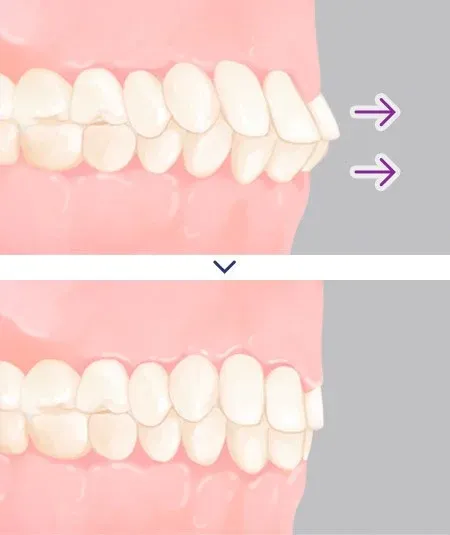
CASE 1. Orthodontic Treatment
For Mouth Protrusion caused by protruding teeth
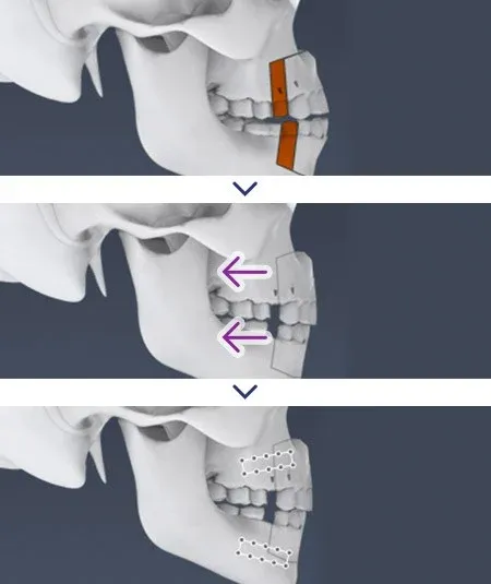
CASE 2. Mouth Protrusion Surgery (ASO)
When both the alveolar bone and teeth are protruding
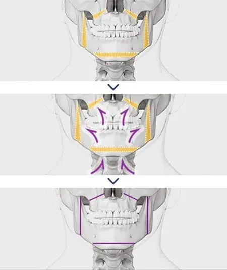
CASE 3. Orthognathic Surgery
For Mouth Protrusion accompanied by malocclusion symptoms
* An accurate treatment plan can be determined after a specialist’s diagnosis based on a detailed examination.
Surgical Method
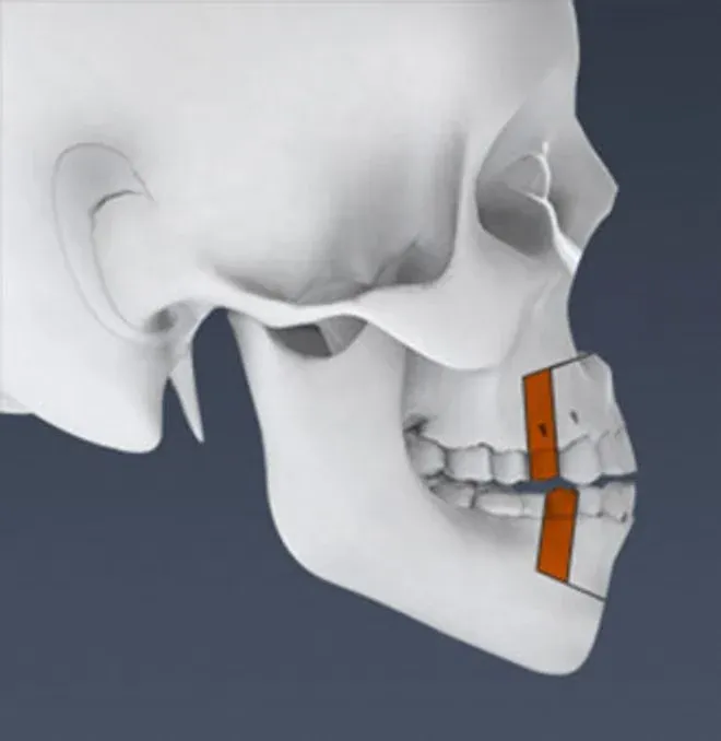
STEP 1. Tooth Extraction and Alveolar Bone Resection
After extracting four upper and lower premolars, the alveolar bone is resected.
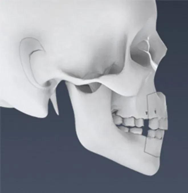
STEP 2. Move the Alveolar Bone and Teeth
The protruding alveolar bone and teeth are moved backward through the extraction spaces.
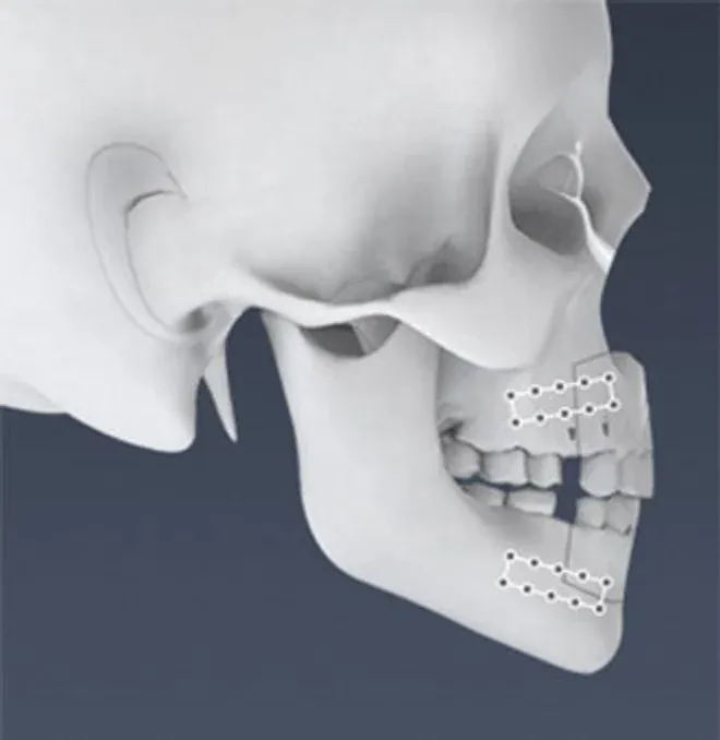
STEP 3. Secure Fixation
The osteotomized bone is firmly fixed in its new position to prevent movement.
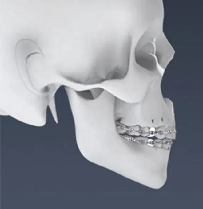
STEP 4. Orthodontic Treatment
Orthodontic Treatment is performed to allow the teeth to settle into position according to the changed bone position.
Surgical Process
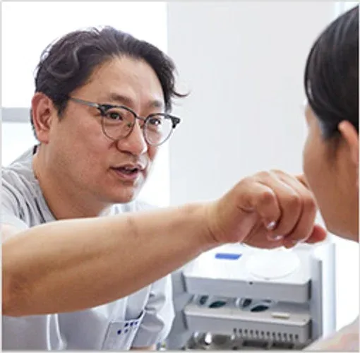
STEP 1. Systematic Consultation
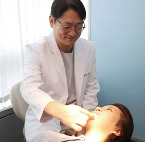
Through a thorough consultation, we identify your desired Face shape and areas for improvement.
STEP 2. Face Shape Analysis
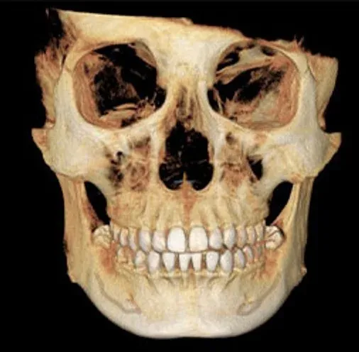
Facial photographs, X-rays, CT scans, and other assessments are analyzed from multiple angles to determine the possible scope of improvement.

STEP 3. Design and Outcome Prediction
Based on the consultation and analysis data, a customized Face shape is designed for each individual.
STEP 4. Safe Surgery
Surgery is performed safely through collaboration among specialists in plastic surgery, anesthesiology and pain medicine, and dentistry.
STEP 5. Aftercare
Progress is monitored regularly with CT and X-rays, along with personalized aftercare.
Features of View Facial Bone Surgery
Point 1. Collaborative Care by Plastic Surgery, Dentistry, and Anesthesiology and Pain Medicine
Facial bone surgery for correcting skeletal malocclusion requires meticulous analysis by specialists with extensive surgical experience. Specialists in plastic surgery, dentistry, and anesthesiology and pain medicine collaborate in treatment.
Point 2. Precision Analysis with 3D CT, X-RAY, and Morpheus
High-resolution diagnostic equipment is used to analyze facial characteristics, dental occlusion, bone shape and size, nerve pathway locations, and the characteristics of muscle and fatty tissue to carefully establish a surgical plan.
Point 3. Use of Ultrasonic Osteotomy Equipment (BoneScalpel)
Ultrasonic bone-cutting equipment is used to reduce damage to surrounding tissues and cut bone with greater precision.
Safety-First System
· On-site anesthesiology and pain medicine specialists and dedicated one-on-one anesthesia care · Special treatment medication for malignant hyperthermia (dantrolene) available for early emergency response · Named surgeon accountability from consultation through surgery and follow-up · In-house inpatient ward · One-on-one rounds by the operating surgeon and 365-day on-site professional nursing staff
Before-and-After Photos of Facial Bone Surgery
These are cases of View Orthognathic Surgery and Mouth Protrusion surgery.

Before surgery → 10 months after surgery

Before surgery → 6 months after surgery
Recovery Progress 1
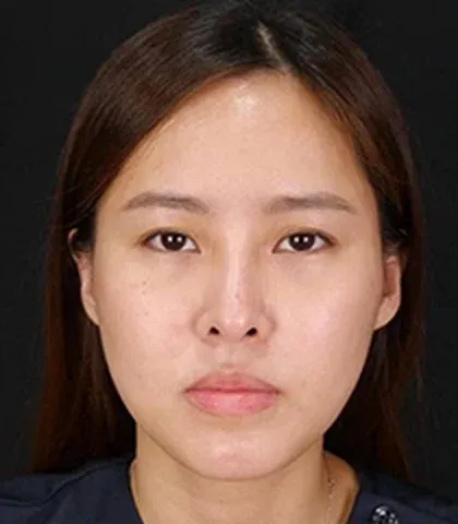
Before surgery

4 months after surgery

5 months after surgery

8 months after surgery
Recovery Progress 2

Before surgery

2 weeks after surgery

2 months after surgery

5 months after surgery
Surgery Information
| Surgery duration: 2 hours | Anesthesia: General anesthesia | Hospital stay: 2 days | Follow-up visits: Approximately 2 visits | Return to daily activities: Approximately 14 days later
Postoperative Care
· 3 days after surgery: Disinfection and progress check, shampoo care · 1 week after surgery: Cleansing and exfoliation; care to support Skin Regeneration and reduce swelling; follow-up CT scan · 2 weeks after surgery: Cleansing; radiofrequency or ultrasound care to support recovery and reduce swelling · 1–6 months after surgery: Regular progress checks Hyperbaric oxygen chamber therapy, medication prescriptions and gargling, anti-swelling injections, Omega Light care, and other treatments may be provided together. After 3D CT and X-ray imaging, the dedicated attending physician reviews progress one-on-one. * The care program and CT scan schedule may change depending on the individual’s condition.
Please Be Sure to Check
The before-and-after photos were taken and published with the patient’s consent. Surgical results and recovery periods may vary depending on individual circumstances, and the surgical method is determined through consultation with a specialist.
 VIEW PLASTIC SURGERY