
Implant Complications (Show-through / Contracture / Migration)
Price upon consultation
Through extensive expertise, we accurately analyze the causes of previous surgical failure to create a nasal contour that harmonizes with the entire face.
Surgery Overview

Brown Rhinoplasty Revision
With skilled expertise, we accurately analyze the causes of unsuccessful outcomes to create a nasal contour that harmonizes with the entire face.
Recommended For
✓ Those whose nose looks unnatural or who are dissatisfied after surgery
✓ Those whose nose has become crooked after surgery
✓ Those who have developed Implant-related problems
✓ Those who have difficulty breathing through the nose after surgery
✓ Cases where the Nasal Tip becomes upturned or the Implant shows through after surgery
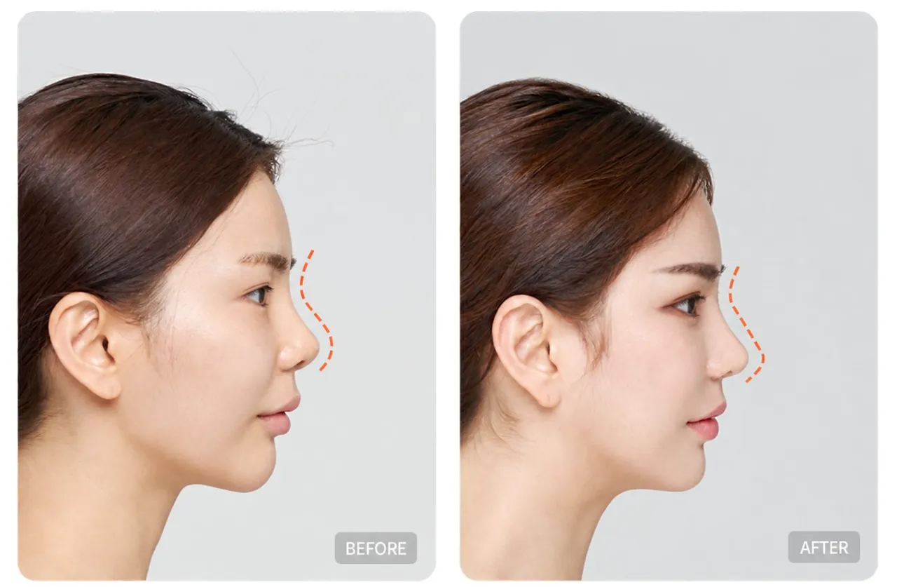
Rhinoplasty Revision to Restore an Unsuccessful Nose into New Beauty
Rhinoplasty Revision refers to a procedure performed again after an initial rhinoplasty when the results are not satisfactory aesthetically or functionally. Compared with the first procedure, revision surgery involves a greater risk of inflammation and tissue damage, requiring a more precise operation. Therefore, it is most important to undergo surgery with thorough analysis and an experienced specialist, and the procedure should be systematically planned with both aesthetic and functional aspects in mind.
01
A more precise operation is required than for the first surgery
02
Stable treatment and surgery performed by an experienced specialist
03
Systematic planning to address aesthetic and functional aspects simultaneously
Rhinoplasty Revision Methods
01
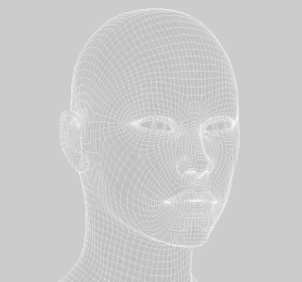
Accurate cause analysis and diagnosis through 3D CT imaging

02
Establishment of a systematic surgical plan

03
Customized nasal shape and angle design suited to each individual's face
04
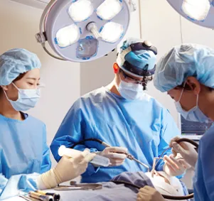
Minimizing tissue damage caused by revision surgery

Key Techniques in Rhinoplasty Revision
Clean capsule removal through extensive experience and skilled technique
Capsule removal is one of the most important techniques for successful Rhinoplasty Revision. A capsule is a membrane that forms when an Implant is inserted into the body. If revision surgery is performed without removing the capsule, it may cause an uneven contour or side effects such as inflammation, so it must be removed. Brown has the extensive experience and skilled techniques needed to remove the capsule cleanly without damaging surrounding tissue.
Types of Rhinoplasty Revision by Case
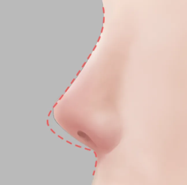
01 / When the Implant Shows Through
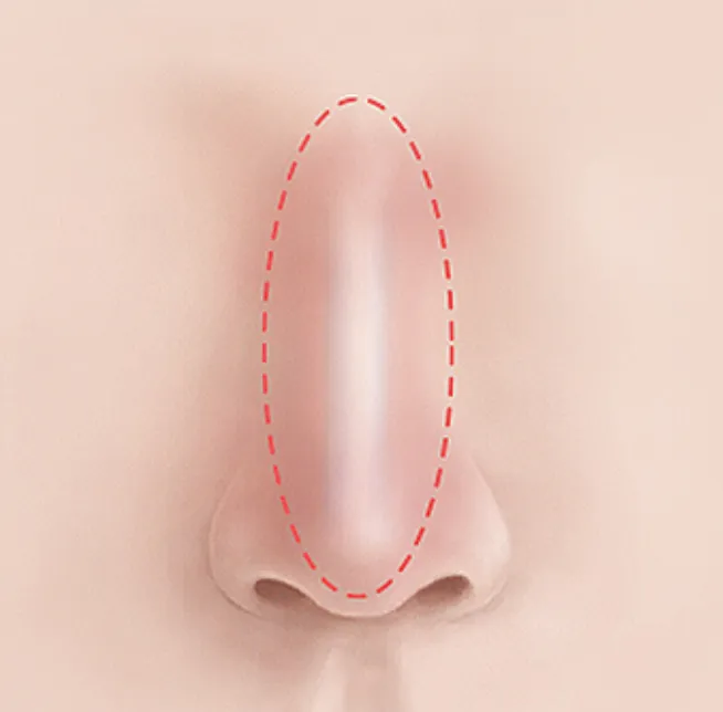
The degree and location of Implant visibility are assessed, and the height is lowered or the Implant is replaced with a thinner one. When the Implant shows through, Dermal Graft or fascia is used to wrap the Implant and correct the visibility.
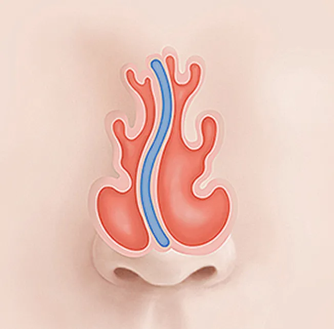
02 / When the Nasal Bridge Appears Crooked
For Rhinoplasty Revision, the existing Implant is completely removed, and asymmetrical skin tissue and facial skeletal structure are assessed before a customized Implant is reinserted. If there is a Septal Cartilage issue, the procedure is performed together with corrective surgery such as Septoplasty.
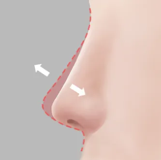
03 / When Capsular contracture or Inflammation Occurs
When localized inflammation occurs, it is treated with antibiotics. When inflammation related to an Implant occurs, the Implant is removed as soon as possible and revision surgery is performed using Autologous Tissue.
04 / When the Nasal Bridge Height Is Unsatisfactory
If the Nasal Bridge is too high or too low after surgery, it is changed and corrected with an appropriate Implant that considers facial proportions and the individual's preferences.
Why Is 3D CT Important?
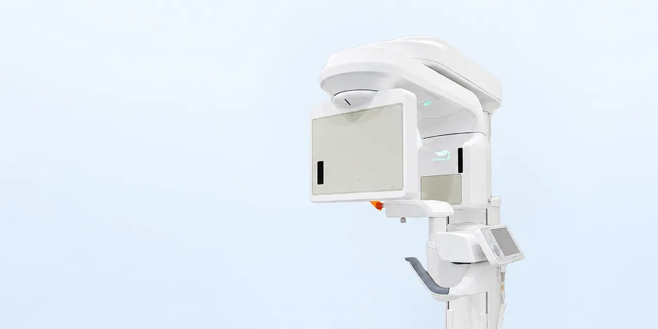
Accurate Diagnosis and Personalized Surgical Planning with 3D CT
At Brown Plastic Surgery, during every rhinoplasty consultation, we use 3D CT to directly examine the nasal skeletal structure, condition of the Septal Cartilage, bone thickness, width, and location. We thoroughly analyze internal nasal issues and establish an appropriate surgical plan for each individual. This allows patients to anticipate predictable, personalized surgical results.
Customized Septal Cartilage Analysis
Analysis of the various shapes and sizes of Septal Cartilage in consideration of each patient's individual characteristics
Treatment for Crooked Nose and Septoplasty
Optimized treatment is provided based on accurate diagnosis of Crooked Nose and Septoplasty
Customized Nasal Structure Adjustment and Improvement
Customized treatment planning through analysis of nasal bone asymmetry and precise width
Detailed Structural Analysis and Surgery
Diagnosis of inferior turbinate asymmetry and Nasal Obstruction Correction to improve the patient's breathing
Various Rhinoplasty Materials
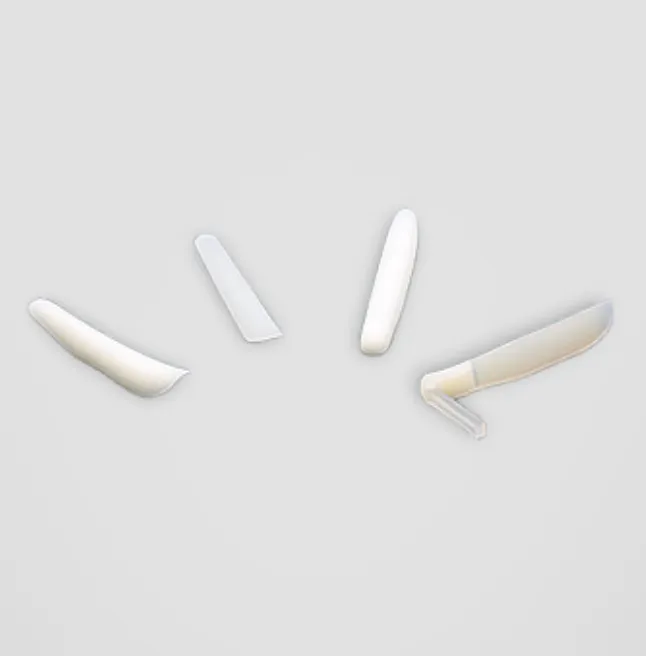
01 / Silicone implant
As the most common Implant, it is known to be relatively safe even when inserted in the body for a long time. It has a low risk of rejection and can be shaped as desired, so it is widely used. Recently, High-soft silicone (Bistool silicone), which feels soft and does not show through, has been developed and is now most commonly used.
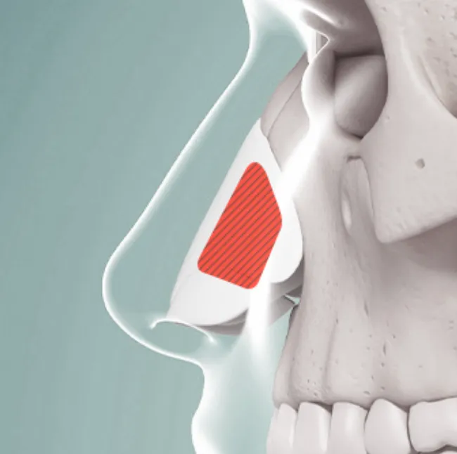
02 / Septal Cartilage
This Cartilage stands like a plate within the partition between the nostrils. Its plate-like shape makes it primarily used to build support for the Nasal Tip.
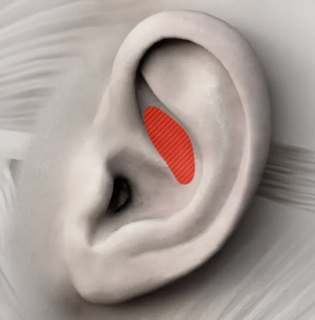
03 / Ear Cartilage
Cartilage from the inner ear is softer than Septal Cartilage and has a curved shape, making it a material that naturally creates the tip shape of the Nasal Tip.
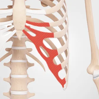
04 / Autologous Rib Cartilage
This method harvests Cartilage from the patient's own breast cartilage for use as an Implant or support structure in rhinoplasty. It is harvested through a minimal incision of approximately 2 cm at the Inframammary fold, most commonly from the sixth to tenth ribs.
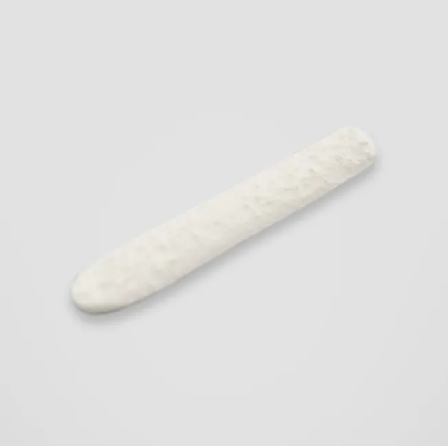
05 / Donor Rib Cartilage
This is breast cartilage obtained from another person's cadaver rather than the patient's own breast cartilage. Cartilage cells between the cartilage material are removed, and radiation treatment is used to eliminate immune reactions before use.
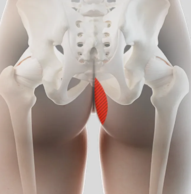
06 / Autologous Dermis
Autologous dermis rhinoplasty uses tissue harvested from the patient's own body rather than a predetermined artificial Implant commonly used in rhinoplasty.
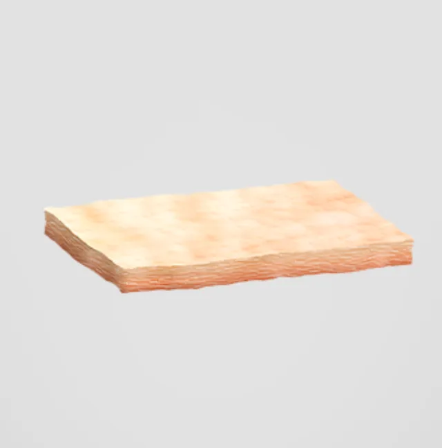
07 / Donor Dermis
This material is made by harvesting another person's skin and removing the epidermis, hair, hair follicles, sweat glands, moisture, and cells, leaving only the dermis.
Before & After Photos
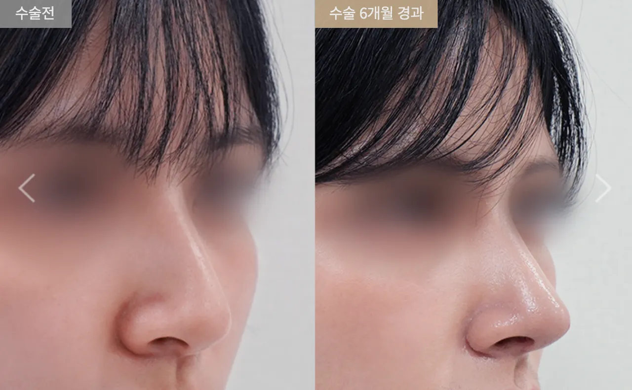
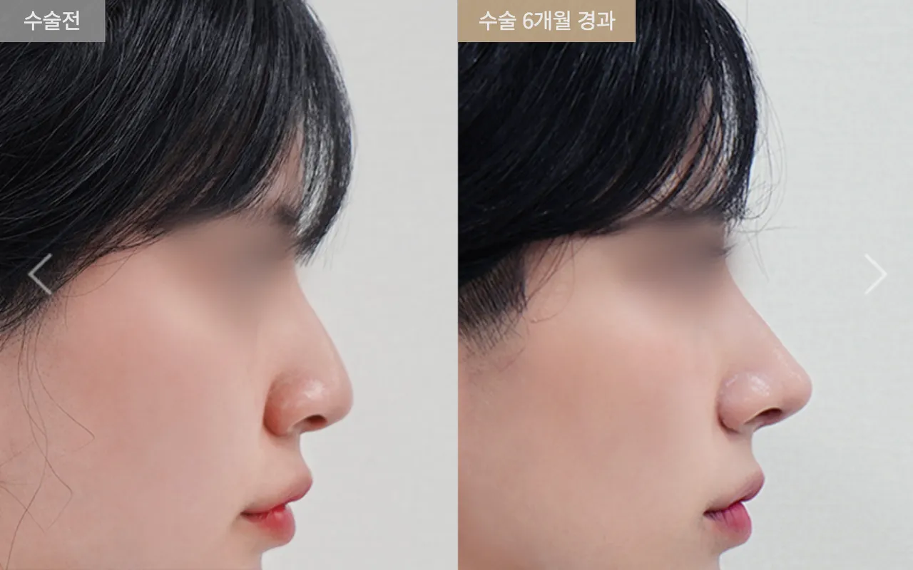
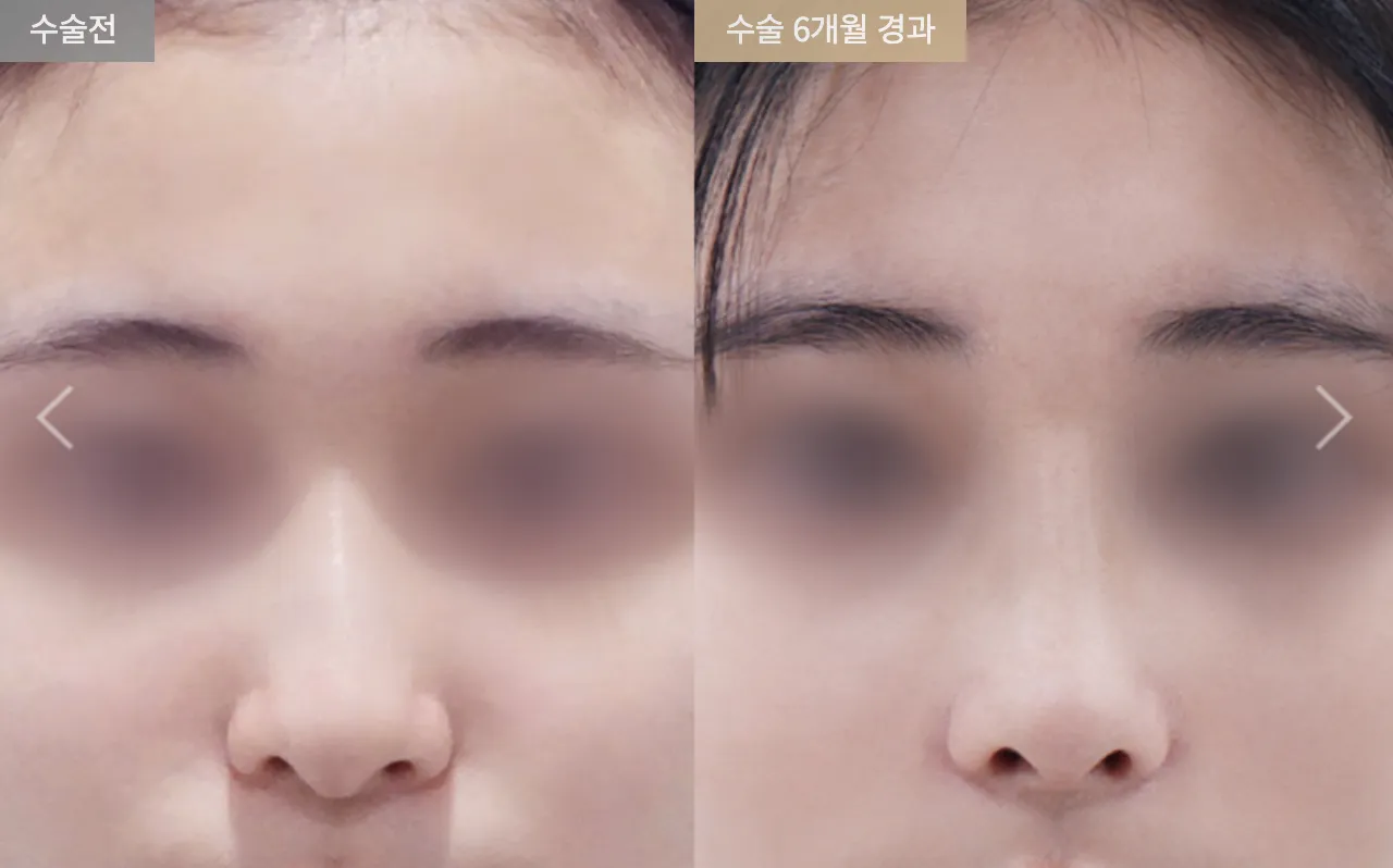
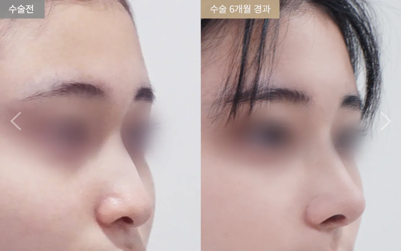
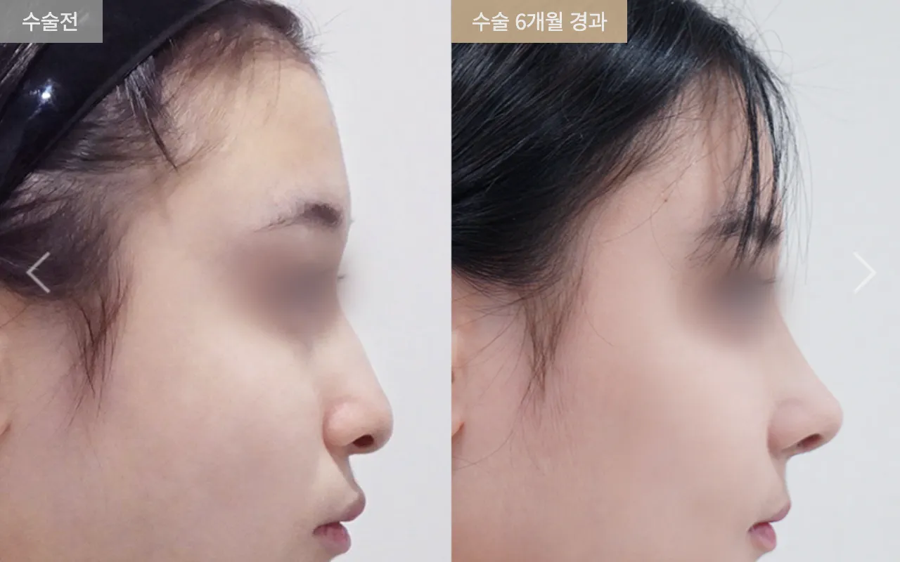
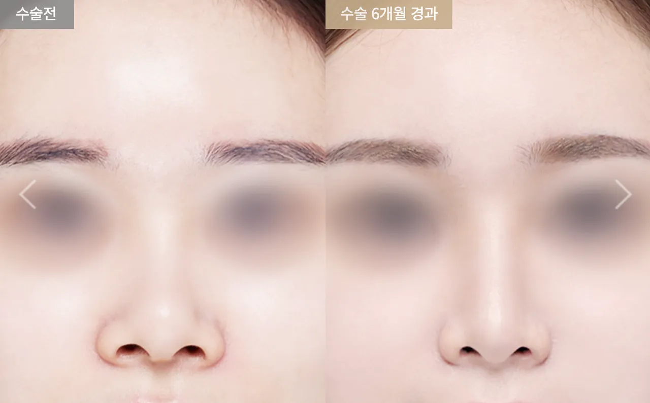
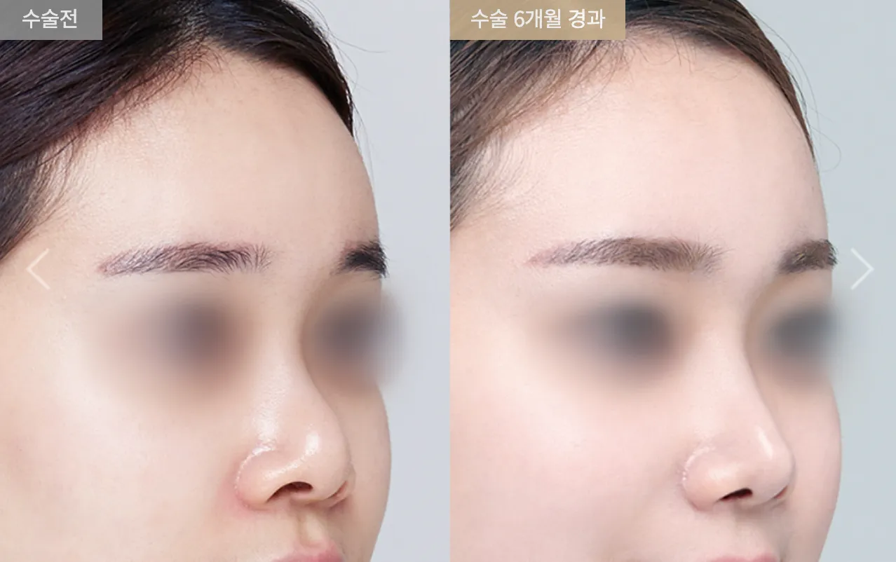
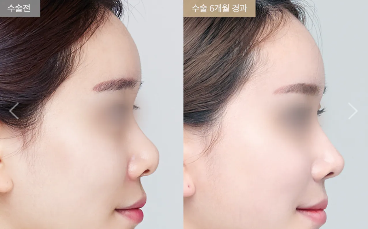
 BRAUN PLASTIC SURGERY
Medical Staffs
 Chief Medical OfficerTaekyu Kim
00