
Glaucoma
Price upon consultation
Glaucoma is an eye disease that damages the optic nerve, which sends visual information to the brain, causing the field of vision to gradually narrow. Since damaged optic nerves are difficult to restore, early detection and ongoing management are important.
Glaucoma
Glaucoma: by preventing progression, you can protect your vision for life.
The leading cause of blindness, but its progression can be controlled and prevented through early detection and consistent treatment.
Definition of Glaucoma
What Is Glaucoma?
Glaucoma is a condition in which the optic nerve, which transmits visual information received by the eye to the brain, is damaged, causing the visual field to gradually narrow. It is not simply a matter of declining vision; because a damaged optic nerve cannot be restored, early detection and management are especially important.
Normal Eye
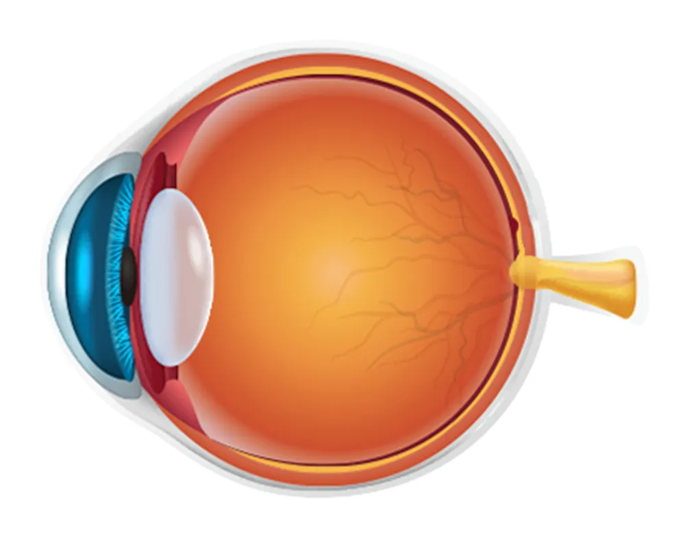
Normal optic disc
Glaucoma
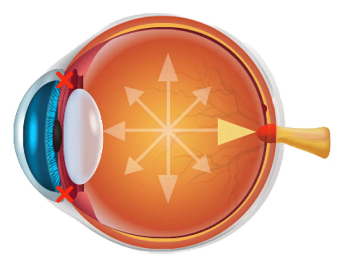
Damaged, enlarged optic disc
Why Is Glaucoma Called the Silent Blinding Disease?

The most significant feature of glaucoma is that there are almost no symptoms in its early stages. Many patients do not notice the gradual narrowing of their visual field and only become aware of a problem after the disease has progressed considerably. In particular, because peripheral vision is gradually lost first, it may seem as though there is no problem in everyday life, even though optic nerve damage may already be progressing. This is why glaucoma is often called a “silent thief of sight.”
Could I Be at Risk for Glaucoma?
Diagnosis According to the Stage of Glaucoma Progression
❶ Myopia and High Myopia
When the eyeball becomes elongated, the structures supporting the optic nerve may weaken. The risk of glaucoma is approximately 1.5–2.3 times higher with myopia and 3.3–4.1 times higher with high myopia.
❷ Thin Cornea
If the cornea is thin, intraocular pressure measurements may be lower than the actual value, requiring caution. The risk increases by approximately 1.3 times for every 40 μm decrease in corneal thickness.
❸ Family History of Glaucoma
Genetic factors may be involved when a parent or sibling has glaucoma. The risk is approximately 1.12–3.69 times higher than in those without a family history.
❹ High Intraocular Pressure
High intraocular pressure increases the likelihood of optic nerve damage due to mechanical pressure. Ocular hypertension is the strongest risk factor, increasing the risk of glaucoma by approximately 13 times.
❺ Impaired Blood Flow Regulation and Systemic Diseases
When blood circulation to the optic nerve is unstable, nerve damage can occur more easily. Conditions include diabetes, hypertension (or hypotension), migraines, hypothyroidism, and sleep apnea.
❻ Increased Cup-to-Disc Ratio (C/D Ratio)
This refers to a large proportion of cupping (the depressed central area) in the optic nerve. The larger the ratio, the greater the likelihood of glaucomatous damage, with risk increasing by approximately 2.12–7.9 times.
❼ Optic Disc Hemorrhage
This refers to small hemorrhages observed around the optic nerve and is an important sign suggesting that glaucoma is currently progressing. In such cases, the risk of glaucoma onset and progression is very high, approximately 5.6–14 times greater.
❽ Retinal Nerve Fiber Layer Changes and Visual Field Defects
This refers to cases in which detailed testing (OCT/visual field testing) suggests thinning of the optic nerve or visual field abnormalities. The risk of progression to glaucoma is approximately 1.3–3 times higher.
❾ Other Ocular Factors
Uveitis: approximately 2–10 times increased risk Pseudoexfoliation syndrome: approximately 5–10 times increased risk Narrow anterior chamber angle: approximately 1.5–3 times increased risk
Glaucoma Self-Assessment
A Glaucoma Self-Assessment You Can Check with Your Own Eyes

The standard for diagnosing glaucoma remains the conventional “standard visual field test.” However, it has been difficult for patients to intuitively understand their own condition through this standard test. The glaucoma awareness test is a proprietary technology invented and patented directly by Gangnam St. Mary's Eye Clinic to address this limitation. Rather than simply pressing buttons as instructed by a machine, this specialized examination—available only at our clinic—is designed to allow patients to directly find and visually confirm their own scotomas (areas they cannot see) on the screen.
Diagnosis According to the Stage of Glaucoma Progression
Even with the same type of glaucoma, the stage of progression differs.
❶ Ocular Hypertension
Intraocular pressure is higher than the normal range (10–21 mmHg), but there is not yet any optic nerve damage. Eye drop treatment may be needed to reduce the risk of progression to glaucoma.
❷ Glaucoma Suspect
This is a condition in which glaucoma is not currently present, but there are risk factors that may lead to glaucoma in the future. The optic nerve may appear suspicious or intraocular pressure may be high, but the condition has not yet been definitively diagnosed.
❸ Normal-Tension Glaucoma
Intraocular pressure is within the normal range (10–21 mmHg), but optic nerve damage progresses because the optic nerve is vulnerable or blood flow is insufficient. This is the most common type among Koreans.
❹ Primary Open-Angle Glaucoma
This type occurs when the eye’s drainage system (trabecular meshwork) gradually loses function, causing intraocular pressure to rise above the normal range and damaging the optic nerve. It is the most common type among Western populations.
❺ Angle-Closure Glaucoma
This condition occurs when the drainage pathway (angle) between the iris and cornea becomes narrowed or blocked, preventing fluid drainage and potentially causing a rapid rise in intraocular pressure. Optic nerve damage may progress rapidly depending on the elevated intraocular pressure. Preventing acute attacks is important.
❻ Pseudoexfoliative Glaucoma
This condition involves dandruff-like white material in the eye blocking the drainage system. Intraocular pressure tends to be higher and progression faster than in typical glaucoma, so caution is needed.
❼ Secondary Glaucoma
This is glaucoma that develops secondary to elevated intraocular pressure caused by other factors, such as uveitis (inflammation), diabetes, trauma, or steroid use. Treating the underlying condition is also important.
Glaucoma Treatment Methods
Glaucoma Treatment Strategies Tailored to Your Individual Condition

Eye Drop Treatment
This is the first and most fundamental step in glaucoma treatment. Think of the eye as a “sink.” Intraocular pressure is safely lowered either by widening the drain so fluid can flow out more easily or by partially turning off the faucet to reduce the amount of fluid (aqueous humor).
Laser Treatment
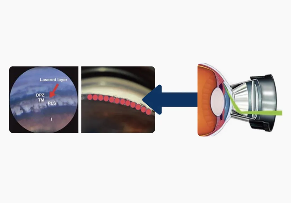
Laser treatment is not hot or painful. A special laser with a very short wavelength is used to gently clean or stimulate cells in the eye’s blocked drainage system (trabecular meshwork), helping restore its drainage function. Because it does not burn or destroy tissue, it is a safe first-line treatment that does not leave structural changes after the procedure.
CPC Laser Treatment
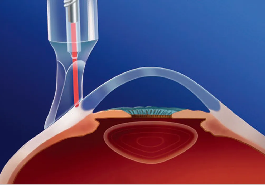
When eye drops alone are insufficient and surgery feels burdensome, this is a non-incisional procedure in which laser energy is applied from outside the eye to gently reduce the function of the ciliary body inside the eye. CPC laser is specialized equipment that has been introduced only on a limited basis even at some university hospitals in Korea.
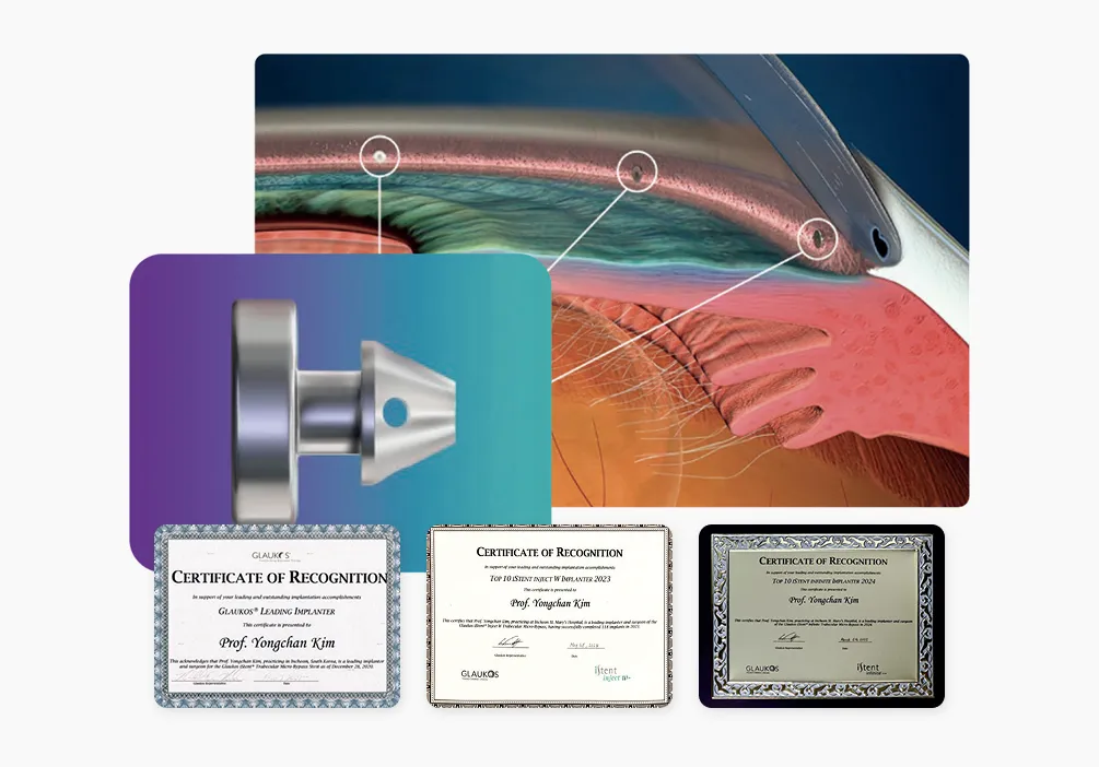
Surgical Treatment: iStent
This is an innovative procedure that performs cataract surgery and glaucoma surgery at the same time. An ultraminiature 0.36 mm stent made of biocompatible medical-grade titanium is implanted into the blocked trabecular drainage system. It lowers intraocular pressure by creating a new shortcut (bypass) through which aqueous humor can drain freely.
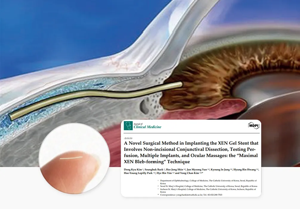
Surgical Treatment: XEN
This procedure implants a microtube made of biocompatible gelatin into the eye, creating a “new pathway” for fluid to drain in place of the trabecular meshwork, whose function has declined. However, using the same material (XEN) does not necessarily produce the same results. Based on experience performing approximately 400 procedures—among the highest volumes in Korea—and proprietary patented technology, Gangnam St. Mary's Eye Clinic delivers optimal results that overcome the limitations of conventional surgery.
 Gangnam Sungmowon Eye Clinic
Medical Staffs
 Chief Medical OfficerYong Chan Kim
00